
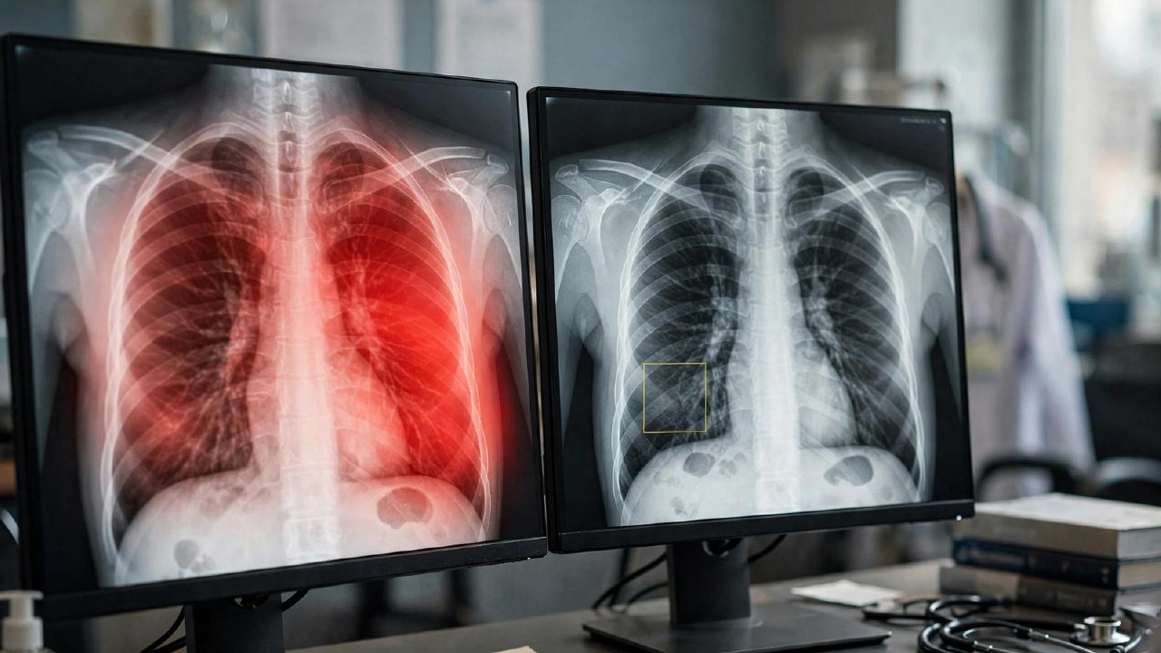
Researchers from the International Institute of Information Technology Hyderabad (IIIT Hyderabad) conducted an audit of four computer vision models designed for chest X-ray analysis. The study evaluated MAIRA-2, MedGemma-4B, LLaVA-Med-1.5, and LLaVA-1.5 by comparing their attention heatmaps against radiologist annotations across three public datasets and a specific pneumothorax patient sample.
The automated comparison ranked MAIRA-2 as the most accurate, followed by MedGemma and the two LLaVA versions. However, the study revealed that in typical cases, the models performed no better than simple anatomical templates based on average data. The AI often highlighted regions where pathologies are statistically common rather than identifying the actual location of the condition on a specific image.
Only in atypical cases, where findings deviated from standard patterns, did MAIRA-2 and MedGemma show localization tied to the specific image. A parallel experiment involving radiologists reviewing 124 cases confirmed that human experts evaluate heatmap plausibility differently than automated metrics, often preferring broader, clinically relevant context over strict adherence to statistical windows.
The authors suggest implementing metrics that account for anatomical prior distributions. They also emphasize the necessity of involving radiologists in the evaluation process for medical AI systems.