

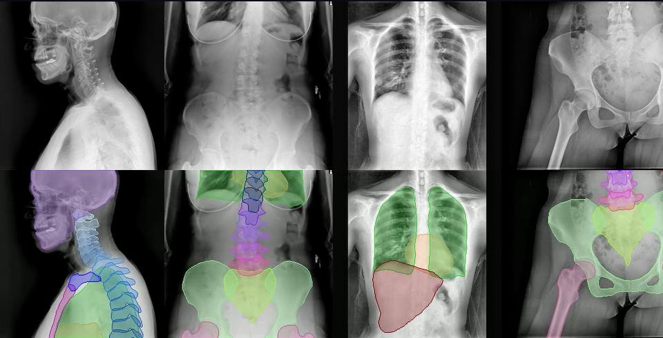
Researchers from MIT, Massachusetts General Hospital, and Harvard Medical School have developed FleXray, an AI model designed to identify 60 anatomical structures in X-ray images. The system supports various projections, patient ages, and imaging types, including standard radiographs and real-time fluoroscopy.
During testing on 213 fluoroscopy images, integrating FleXray with CT alignment algorithms reduced the median error from 335 mm to 1.2 mm. Additionally, the rate of unsuccessful alignment attempts dropped from 86% to 13%. The model utilizes a U-Net convolutional neural network architecture and can combine predictions from five separate models to enhance accuracy.
The model achieved a mean Dice coefficient of 0.875 across eight independent datasets. It outperformed specialized models like PAXray and FluoroSAM in seven of those sets. While specialized nnU-Net models achieved a higher score of 0.914, FleXray demonstrated strong versatility in tasks such as measuring the Cobb angle for scoliosis, where it achieved a mean absolute error of 5.13 degrees.
To train the model, researchers used synthetic X-rays generated from CT scans, enhanced with realistic noise and exposure variations via the FLUX.2 image editing model. FleXray also supports transfer learning for tasks like pediatric pelvic segmentation and bone tumor detection. The software runs locally on user devices via WebGPU or WebAssembly.